Parts Of An Animal Cell Visible Under A Light Microscope / Muppets Animal Drawing at PaintingValley.com | Explore ... : As for seeing electrons under any microscope in general.
byEilene Pierceall-
0
Parts Of An Animal Cell Visible Under A Light Microscope / Muppets Animal Drawing at PaintingValley.com | Explore ... : As for seeing electrons under any microscope in general.. See how a generalized structure of an animal cell and plant cell look with labeled diagrams. An animal cell also contains a cell membrane to keep all the organelles and cytoplasm contained, but it lacks a cell wall. The animal cell is more. Which part of the compound microscope helps in gathering and focusing light rays on the specimen to be answer: Under a microscope, plant cells from the same source will have a uniform size and shape.
Diagram 3.2 an animal cell. The ability of a microscope to distinguish two adjacent points as distinct and. Electron microscopes use accelerated electron beams (as opposed to visible light in a light microscope) to create images of magnification as high as 1 million x and has a very high resolving power to see the objects in fine detail. An animal cell also contains a cell membrane to keep all the organelles and cytoplasm contained, but it lacks a cell wall. As per the given information in the question, cells of mushrooms, plants, and animals all have visible nuclei under a microscope.
Electron Microscope Eukaryotic Animal Cell - Micropedia from image2.slideserve.com As per the given information in the question, cells of mushrooms, plants, and animals all have visible nuclei under a microscope. Which of the following cell structures can you see under a light microscope? Cell is a tiny structure and functional unit of a living organism containing various parts known as organelles. As for seeing electrons under any microscope in general. The organelles in a plant cell vary in size. Observing a wide range of biological processes and animal cell under light microscope is easier due to advances in microscopic techniques. (b)(i)state which of the following organs has the highest number of mitochondria per cell: Microscopes and how to use a light microscope.
Cell organelles explained in 5 minutes!!
Beneath a plant cell's cell wall is a cell membrane. Plant and animal cells can be studied in greater detail with a light microscope by magnifying the image. Magnifying is the purpose of a microscope and thus used observe a thing or organisms which are too tiny to see with unaided eye. The colors of a prepared tissue are not natural colors. What can only be seen under a microscope can now cover an entire serving plate. Dna is a very important part of all cells and therefore of all life. An animal cell also contains a cell membrane to keep all the organelles and cytoplasm contained, but it lacks a cell wall. Mitochondrion b explain why ribosomes are not visible using a light microscope. Cell is a tiny structure and functional unit of a living organism containing various parts known as organelles. Resolution of a microscope is. Under a microscope, plant cells from the same source will have a uniform size and shape. The organelles in a plant cell vary in size. (i)mirror (ii)eye piece lens (iii)fine adjustment knob.
Beneath a plant cell's cell wall is a cell membrane. Dna contains information that encodes all our. (i)mirror (ii)eye piece lens (iii)fine adjustment knob. (ii)give your answers in b state the function of each of the following parts of a light microscope: Animal cells also have a many of the differences between plant and animal cells are visible under a microscope, and it's relatively straightforward to distinguish between the two.
What Are the Differences Between a Plant & an Animal Cell ... from img-aws.ehowcdn.com To examine objects using a light microscope (light microscopy), it is necessary to be able to (a)how is mitochondria adapted to its function? What can only be seen under a microscope can now cover an entire serving plate. Some features common to animal cells. As for seeing electrons under any microscope in general. Smooth endoplasmic reticulum is found in both animal and plant cells and it serves different functions in each. Resolution of a microscope is. The cell membrane is selectively permeable in nature, consisting of a lipid bilayer with proteins, glycolipids, and cholesterol attached to them in a specific pattern.
The organelles in a plant cell vary in size.
Although cells are diverse, all cells have certain parts in common. Cells consist of cytoplasm enclosed within a membrane, which contains many biomolecules such as proteins and nucleic acids.2 most plant and animal cells are only visible under a light microscope, with dimensions between 1 and 100 micrometres.3 electron microscopy gives a much higher. The cell membrane is selectively permeable in nature, consisting of a lipid bilayer with proteins, glycolipids, and cholesterol attached to them in a specific pattern. You see that many features are in common. The compound microscope is a precision instrument. Mitochondrion b explain why ribosomes are not visible using a light microscope. As per the given information in the question, cells of mushrooms, plants, and animals all have visible nuclei under a microscope. Cell is a tiny structure and functional unit of a living organism containing various parts known as organelles. Plant cells have cell walls, one large vacuole per cell, and chloroplasts, while animal cells will have a cell membrane only. Observing a wide range of biological processes and animal cell under light microscope is easier due to advances in microscopic techniques. The advancement of light microscopy also required methods for preserving plant and animal tissues and making their cellular details more visible, methods the slices of tissue, called histological sections, are typically thinner than a single cell. The ability of a microscope to distinguish two adjacent points as distinct and. In the above observation of onion cells, you can see the cell surface as you can see in the above labeled plant cell diagram under light microscope, there are.
Resolution of a microscope is. (a)how is mitochondria adapted to its function? A generalised animal cell as observed under an electron microscope. Which of the following cell structures can you see under a light microscope? As for seeing electrons under any microscope in general.
Epidermal onion cells under a microscope. Plant cells ... from i.pinimg.com The compound microscope is a precision instrument. The animal cell is more. Microscopes using the visible part of the electromagnetic spectrum (visible light) were invented in the 16th century and the. These are all common parts of a cell. Dna contains information that encodes all our. Dna is a very important part of all cells and therefore of all life. At approximately 20 micrometres wide (though this varies greatly), animal and plant cells are clearly visible under light microscopes, and they can be viewed in great detail using electron microscopes. However, they usually can achieve a maximum of 2000x magnification which is not sufficient to see many other tiny organelles like ribosomes, endoplasmic reticulum, lysosomes, centrioles, golgi bodies unless they have an electron.
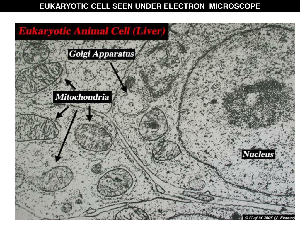
Under a light microscope, the parts of a simple animal cell (e.g.
We say cells are microscopic because they can only be seen under a microscope. Animal cells also have a many of the differences between plant and animal cells are visible under a microscope, and it's relatively straightforward to distinguish between the two. Learn about and revise cell structures with bbc bitesize for gcse biology, ocr gateway. Faintly visible are several mitochondria, for example the grey oval structures at the bottom left. In most plant cells, the organelles that are visible under a compound {light} microscope are the cell wall, cell membrane, cytoplasm, central vacuole here is an electron micrograph of an animal cell with the labels superimposed: Some features common to animal cells. The compound microscope is a precision instrument. Under the microscope, an animal cell shows many different parts called organelles, that work together to keep the cell functional. Below the basic structure is shown in the same animal cell, on the left viewed with the light microscope, and on the right with the mitochondria are visible with the light microscope but can't be seen in detail. A generalised animal cell as observed under an electron microscope. As per the given information in the question, cells of mushrooms, plants, and animals all have visible nuclei under a microscope. Most cells, both animal and plant, range in size between 1 and 100 micrometers and are thus visible only with the aid of a microscope. A compound light microscopes use lenses and light to magnify cell parts.